|
Motivation: |
|
Project Description:
We chose to couple liquid phase IEF to
free solution electrophoreiss. Ampholytes were used
as the running buffer for both dimensions, while boundary condition control
of the applied voltage and terminal buffer chemistries was used to govern the
acting separation mechanisms. Low-dispersion electroosmotic flow mobilized
focused species to junctions in a t-geometry. Once species arrived at
the junction, they were electrokinetically-sampled
into the electrophoresis dimension. Subsequently, species remaining in the
IEF dimension were refocused and subjected to electrophoretic analysis. This
algorithm was repeated until all fluid volumes of interest from the first dimesnion were analyzed by the second dimension. We have shown that on-chip IEF acts to
focus an initially uniform sample into a tight volume at the isoelectric
point (pI) resulting in enrichment factors of 80x Species focused
quickly in the IEF dimension (< 60s) and can be rapidly sampled and
analyzed by the second dimension (periods of 1 min). Two-dimensional
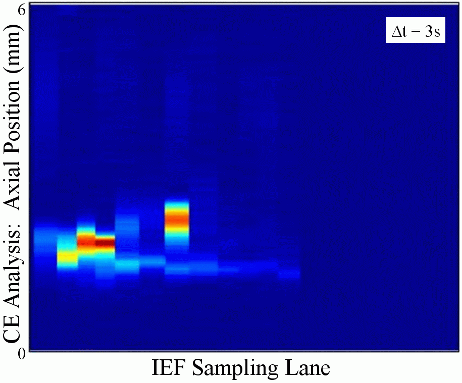
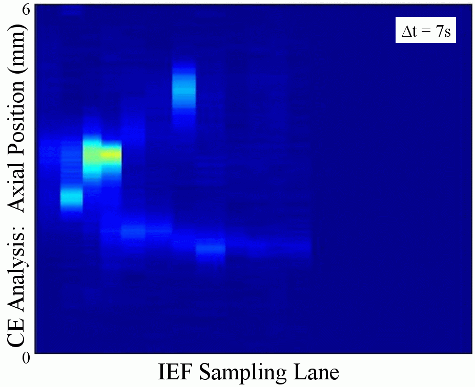
gel-like plots were constructed from time-sequences of CCD images collected
during the 2D separations (Figure at right). The gel-like plots were
formatted to display intensity information so as to mimic a slab-gel
result. Based on the CCD images, the horizontal axis of the gel-like
plots represents the relative location of the fluidic volumes during
IEF. The vertical axis of the gel-like plots corresponds to the axial
spatial coordinates of the second dimension. Thus, each vertical lane
of the 2D separation corresponds to successive CE separations at the same
elapsed CE analysis time. |
|
|
References:
Collaborators: |
aeh
07.24.03